Other Less
Common Causes
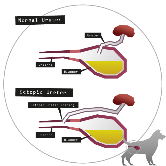
Ectopic Ureter
The definition of ectopic in medical terms is ‘abnormal place or position’. In UI this is when the ureter, which is the tube that connects the kidney to the bladder, joins the bladder in an abnormal place. It should normally join in the main body of the bladder as shown in the first diagram below. If it does not enter here (if not then it often enters into the urethra as shown in the second diagram below) it is termed an ectopic ureter.
Pets with this condition usually have continuous leakage that will have occurred since birth and will often fail to respond to any medication prescribed for USMI or a Urinary tract infection (UTI).
This is more common in female than male animals. The cause is unknown but hereditary factors play a role and although rare, this would be the commonest cause of UI in juvenile dogs1.
The condition is usually diagnosed using special contrast X-ray techniques that use a contrast medium to highlight tissues and organs that would not be visible on a normal plain X-ray.
The only treatment option is surgery to attempt to re-implant the ureter in the correct position.
Bladder Hypoplasia
This basically means that the dog has been born with a bladder which is relatively too small. This is diagnosed by contrast X-ray.
Pervious Urachus
This happens when a communication which is present in the foetus between the bladder and the umbilical cord fails to close before birth. This means that there is a leakage of urine and can lead to scalding of the skin in the tummy region. This is a very rare condition and is diagnosed using contrast X-rays.
Intersexuality
This is when the dog is born with male and female genitalia. Again this condition is very rare and is usually diagnosed with contrast X-rays.
Congenital Neurological Conditions
Another rare condition is when a puppy is born with a problem with the spine. This condition will be quite obvious as the main signs you will spot are an inability to stand and/or walk normally but they may also be incontinent. To diagnose this your vet will need to perform spinal X-rays and/or MRI (Magnetic Resonance Imaging - strong magnetic fields and radio waves form images of the body).
Detrusor Instability
The detrusor muscle is found in the bladder wall and when it contracts it is responsible for emptying the bladder. Detrusor instability is a rare condition that occurs when this muscle is unable to remain relaxed during the urine storage phase.
It usually occurs as a result of persistent bladder wall stimulation which may exist when cystitis (bladder infection) is present but may occur with no underlying cause.
Leakage occurs at irregular intervals and is not continuous or steady. It is more likely to be associated with activity or excitement and happens due to involuntary contractions.
Dogs will often get into position to urinate when stimulated by a detrusor contraction.
This condition is rare in companion animals but it can occur alone or in combination with USMI in male or female dogs. Treatments are available for this condition which either help to increase bladder capacity and decrease spasticity (tightness of muscles).
Prostatic Disease
The prostate gland surrounds the urethra in a male dog. If it gets enlarged or inflamed it can have an effect on the urethral function which can mean bladder control is lost. Your vet can perform tests to diagnose if this is the case.
How is urinary incontinence investigated
*Disclaimer – some of the drugs mentioned on this website may not be available in your country or may be being used off label, please speak to your vet for information.

Share this with your friends
